MIFOBIO 2025
Biological imaging has made a major contribution to our understanding of living organisms, thanks to a succession of conceptual and technological advances. The fact that several Nobel Prizes have been awarded in this field over the last two decades is a marker of this rapid development, particularly in photonic microscopy. These imaging techniques are developing both in nanoscopy, with measurements at the level of the single molecule, and in macroscopy, with the imaging of whole tissues and organisms. This dynamic is being driven by a succession of major advances in physics, chemistry and molecular and cellular biology, as well as in data analysis and artificial intelligence, aimed at achieving holoscopic multi-scale imaging. It is this interdisciplinary approach, aimed at understanding all the mechanisms of living organisms, that is the focus of our project.
The aim of the MiFoBio school is to bring together experts from a wide range of disciplines working in the field of imaging for biology, so that they can exchange and share their interdisciplinary knowledge and expertise. It provides participants with a common base of knowledge and enables them to learn about new technologies in the field, from all the disciplines involved (cell marking, optical techniques, image processing, analysis, etc.), with the aim of eventually leading them to develop new ways of observing and understanding the functional organisation of cells and tissues. Through courses and practical workshops, as well as round-table discussions, the MiFoBio school aims to encourage conceptual and technological breakthroughs in the understanding of the different levels of organisation of living organisms.
This year, Mifobio is hosting the “CA22153 – European Curvature and Biology Network (EuroCurvoBioNet, see website HERE)” action as part of a European COST (European Cooperation in Science and Technology)! You’ll find a list of the 5 sessions organized on the school program page!
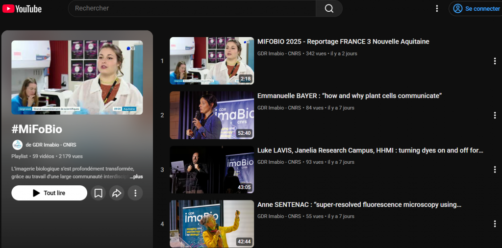
Find all the Mifobio 2025 conferences on our YouTube channel:
Topics covered
Advances in microscopy rely heavily on the ability to create contrasts. The development of specific labelling techniques for molecules or structures of interest also brings added specificity. Various aspects of chemistry have been used to develop labelling techniques adapted to the different scales of organisation of living organisms (cells, tissues, organs and organisms), from the organic or inorganic chemistry of small fluorophores, the chemistry of organic, inorganic or hybrid nano-objects, but also genetically encoded fluorophores, click chemistry in the cell, and biological nano-assemblies such as viruses. The aim of this module is to provide an overview of the various issues involved in cell labelling, from the fundamentals to applications. Biosensor-type tools offering novel access to intracellular information (proximity of proteins, mechanical tension, etc.) will be introduced here and developed at the different organisational scales of living organisms in subsequent modules.
Super-resolution microscopy has become an essential tool in the field of cell biology, making it possible to go beyond the optical diffraction limit and achieve resolutions of up to ten nanometres in 3 spatial dimensions. The challenge is to adapt these techniques for use on living cells and multi-cellular systems. This makes it possible to monitor molecular or cellular processes over time. This opens up new perspectives in the understanding of numerous molecular mechanisms on a nanometric spatial scale and with a temporal resolution of up to a millisecond. This module will provide an overview of the different techniques, highlighting some of the recent developments and the challenges that remain. The aim is to facilitate the democratisation of these approaches by pooling current knowledge.
In the space of a few decades, microscopy has overcome a number of hurdles, both in terms of resolution and functional and tissue imaging. The amount of data acquired and digitised is considerable. Analysing this data has always been a major challenge in biological imaging. Artificial intelligence represents a major change in this field, which has long been dominated by morpho-mathematical approaches and physical analysis models. The algorithms underlying these new analysis techniques are also being used to model results or develop simulation methods. The speed and power of these techniques make it possible to address complex questions and open up new perspectives for deciphering living organisms at different scales. However, is it reasonable to entrust our data analysis to black boxes or statistical analyses? Do powerful tools sometimes make us forget the reality of the physical aspects of our measurements? Or, on the contrary, do they allow us to explore previously inaccessible levels of complexity? But are these tools really that different from Bayesian methods, mathematical approaches or modelling? The aim of this module is to take advantage of the last few years and the numerous implementations of AI approaches to demystify their power and better master their limits. The current challenges are not only to share the knowledge developed in the various laboratories, but also to overcome the obstacles to annotation.
Morphological and functional imaging of organised multicellular systems is a major challenge for a wide range of applications in biology. Their development is highly interdisciplinary: development of new probes, label-free imaging modalities, beam shaping and non-linear optics, optogenetics, etc. In this context, inter-scale imaging is becoming necessary to obtain information on the cell as a whole and on intra-cellular mechanisms at the same time. Organoids and on-chip devices have developed rapidly and now require dedicated imaging tools. All of these strategies will be covered in this module, focusing on tissue imaging and 4D self-organisation of cells.
Understanding the mechanisms that ensure the formation, growth, coherence and adaptation of living organisms is a major challenge. Communication between cells, signalling and the mechanobiological responses of cells are obviously at the heart of the mechanisms being studied. The ambition is to move from a carpet of cells to 3D functional biological structures. This means imaging thick objects (> 100 µm). In addition, optogenetic methods have emerged as extremely relevant tools for controlling and studying target molecular activities. The current challenges are not only to share the knowledge developed in the various laboratories, but also to overcome the obstacles to imaging thick tissue at molecular resolution. These issues are of interest to a wide range of communities working on a variety of themes: mechanotransduction, signalling and regulation, dynamics of regulation and signalling networks, meristems and roots, membrane plasticity, host/parasite relationships, metastasis, embryonic development and regeneration, immune response, wound healing, etc.
This module is offered in collaboration with the GDR Ondes. Its aim is to extend imaging techniques for living organisms by introducing new concepts based on the spatial and temporal control of waves (imaging through highly scattering media), imaging methods using molecular spectroscopy (Raman, IR, autofluorescence, etc.), which are currently being rediscovered, and other non-photonic imaging methods (ultrasound, etc.). One of the major challenges is to improve the penetration of light into tissues by limiting absorption and scattering. To achieve this, various approaches have been implemented, such as non-linear imaging methods using beams shifted towards the infrared or endogenous contrasts, or more recently the use of speckle. The possibility of adapting microscope parameters/performance in real time, as well as the observation modality, is an emerging field (smart microscopy). The main aim here is therefore to present a range of new imaging concepts derived from physics, making it possible to overcome certain current obstacles (mainly depth imaging).
Teaser Mifobio 2023
Mifobio School
MiFoBio is a high-level course combining theoretical and experimental approaches, bringing together academics and industrialists, researchers, engineers and students from different disciplines. It provides an interdisciplinary gateway to functional biology for scientists from different disciplines, in an integrative manner and based on the co-production of knowledge. Its aim is to provide participants from different disciplines with a common base of knowledge and know-how, to enable them to learn about new technologies in the field and to share their skills. MiFoBio is a truly temporary interdisciplinary laboratory offering participants a comprehensive theoretical and practical overview, from biological questions to image processing, from instruments to modelling and analysis.


The MiFoBio school offers a range of courses, round tables and practical workshops organised around 6 themes. In addition to the 70 courses and seminars, 25 round tables and mini-symposia, MiFoBio has an exceptional range of instruments to support a series of workshops run by the participants themselves. More than 130 different workshops will cover the most advanced technologies in the field (ultra-high resolution, light sheet, adaptive optics, non-linear optics, correlative microscopy, etc.). This thematic school offers a genuine temporary interdisciplinary laboratory, including a Fablab, an Optic Lab, or culture room, which provides participants with a complete theoretical and practical overview, from the biological question to the exploitation of the image, via the instrument, right through to modelling and automated analysis. This approach is applied at all levels, and is tailored to each participant’s job or project, just like in a permanent laboratory.
Targeted audience
This school, organised by the members of the Imabio GDR, reaches a much wider audience through calls to other organisations (CEA, INSERM, INRIA, universities, etc.) and learned societies. Any researcher, teacher-researcher, engineer, post-doc or doctoral student from the various disciplines involved in developing or implementing functional microscopy tools is welcome to attend (subject to the number of places available!).
Communication
Find the logos and posters for the 2025 edition of Mifobio.