MIFOBIO 2023


Biological imaging has made a major contribution to our understanding of the living world, thanks to a succession of conceptual and technological advances in chemistry and physics. Over the past 20 years, the field of biological imaging has made dazzling progress, as witnessed by the award of the Nobel Prize in Chemistry in 2008 to O.Shimomura, M. Chalfie and R. Y. Tsien for their work on proteins, in 2014 to E. Betzig, S. Hell and W. Moerner for super-resolution microscopy and finally this year, it was awarded to M. Bawendi, L. Brus and A. Ekimov for the development of Quantum Dots, withmany applications in imaging. These imaging techniques have developed both in nanoscopy, with measurements at the level of the single molecule in a cell, and in macroscopy, with imaging of whole tissues and organisms. Increasingly powerful algorithms make it possible to exploit the vast amounts of data produced by these different imaging techniques.This dynamic is driven by a succession of major advances in mathematics, computer science, physics, chemistry, instrumentation, engineering and molecular and cellular biology. This approach, which aims to integrate increasing levels of complexity, is naturally interdisciplinary. Conceptually and technologically, these imaging technologies need to converge to reintegrate molecular quantification into approaches that take account of physiological aspects on the scale of organs and organisms, whether natural or reconstructed. This means rethinking our imaging methods, exploiting new emerging concepts in physics and chemistry, and taking advantage of new experimental and theoretical models to address issues of greater complexity, integrating notions of biological systems and the effects of their environment. More integrative and systemic approaches combining measurements, analyses and modeling at different spatial and temporal scales of living organisms are needed today. The challenge for MiFoBio school is to share the knowledge and know-how needed to forge new tools and concepts to meet these challenges.

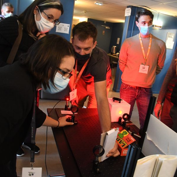
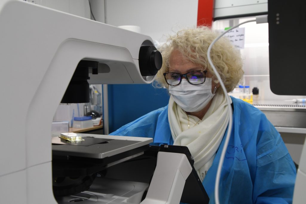
New developments in bioimaging are continuously breaking barriers in terms of resolution, speed, minimal invasiveness, and adaptability to molecular studies in living organisms. As such, the main mission of the MiFoBio school is to ensure the knowledge transfer within the community, training the younger generations at the bioimaging interface in the plurality of its components while also promoting the integration of new skills, particularly in the field of analysis, data management and modeling.

Access program


Get the Web app Mifobio 2023

Essential information about Mifobio can be found in the Mifobio web app, which can be accessed from any mobile phone by scanning the enclosed QR-code.

To access the computer version, follow this link:

https://thunkable.site/w/wQuH9J7MCNubm19Y3ysL_

QR-code to scan to access to the web app

Mifobio TEASER 2023

mifobio SCHOOL

MiFoBio is a high-level training program combining theoretical and experimental approaches, bringing together academics and industrialists, researchers, engineers and students from different disciplines. It provides an interdisciplinary gateway to functional biology for scientists from different disciplines, in an integrative, co-production mode. Its aim is to provide participants from different disciplines with a common base of knowledge and know-how, to enable them to learn about new technologies in the field, and to share their skills. MiFoBio is a veritable interdisciplinary laboratory, offering participants a complete theoretical and practical overview, from biological questions to image processing, from instruments to modeling and analysis.

Last settings on a "homemade" microscope. Credit: Serge Monneret
A cell culture room is set up for the duration of the Mifobio school. Credit: Serge Monneret

The MiFoBio school offers a range of courses, round-table discussions and practical workshops organized around 6 themes. In addition to 70 courses and seminars, 25 roundtables and mini-symposia, MiFoBio boasts an exceptional range of instruments to support a series of workshops run by the participants themselves. Over 130 different workshops will cover the most advanced technologies in the field (ultra-high resolution, light sheet, adaptive optics, non-linear optics, correlative microscopy, etc.). This thematic school offers a truly temporary interdisciplinary laboratory, including a Fablab, an Optic Lab, or culture room, providing participants with a comprehensive vision that ranges from the biological question to image processing, via the instrument itself, right through to modeling and automated analysis. This approach is applied at all levels, and is tailored to each participant’s profession or project, just as in a permanent laboratory.

2023 program

Axes du programme – 10ème édition

– De la molécule à la microscopie : Stratégies de marquage, sondes et contrastes

– Quantification à l’échelle nanométrique et études des assemblages supra-moléculaires

– Dynamiques et interactions moléculaires en cellule et tissus : expérimentation et modélisation

– Ondes sur le vivant : Les nouvelles imageries des organismes

– Imageries des systèmes biologiques auto-organisés, de la cellule à l’organe

– Imagerie et mesures physiques, mécanobiologie- De la molécule à la microscopie : Stratégies de marquage, sondes et contrastes

partners

Testimonials


Public

This school, organized by the members of the GDR ImaBio, reaches a much wider public through calls to other organizations (CEA, INSERM, INRIA, universities… ) and academic societies. Any researcher, lecturer, engineer, post-doc or PhD student from different disciplines involved in the development or implementation of functional microscopy tools is welcome (subject to the available number of places!).